Understanding Your Results

Your screening may identify areas that need a closer look

HerScan provides breast ultrasound screening, not a diagnosis. If your screening identifies an area that requires additional evaluation, your report may recommend following up with your healthcare provider for diagnostic imaging or other testing.

> Jump to Your Result Category

Understanding your HerScan breast ultrasound results

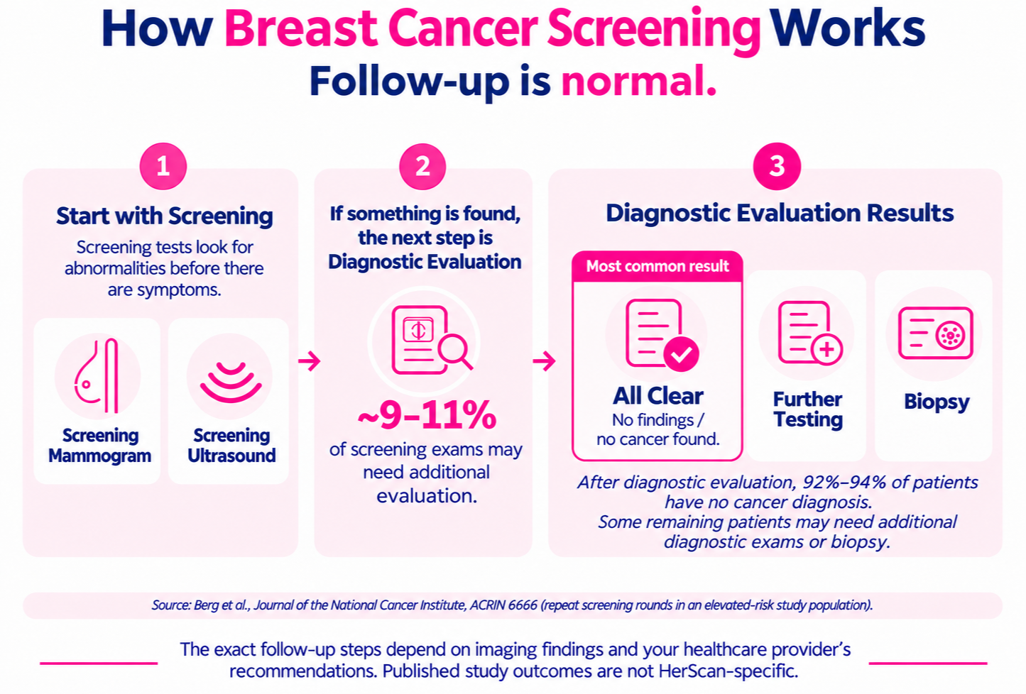
how breast cancer screening works

Screening tests are designed to look for abnormalities in people who may not have symptoms. If a screening identifies something that needs a closer look, additional diagnostic imaging may be recommended. Depending on those results, additional testing or a biopsy may sometimes be needed.

HerScan provides screening breast ultrasound, which is different from diagnostic ultrasound. Diagnostic ultrasound is a targeted exam used to evaluate a specific finding or concern.

Learn More: Screening Ultrasound vs. Diagnostic →


Your Results at a Glance

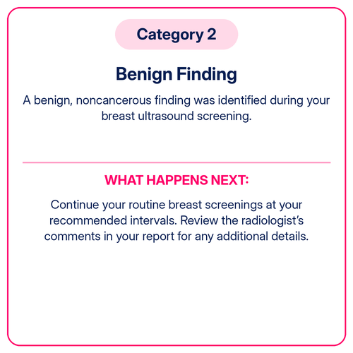
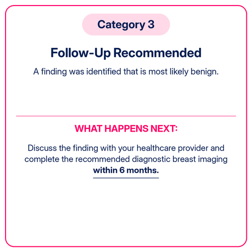
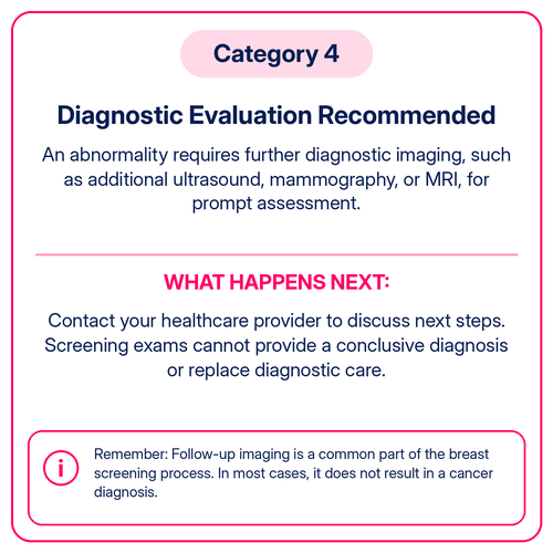
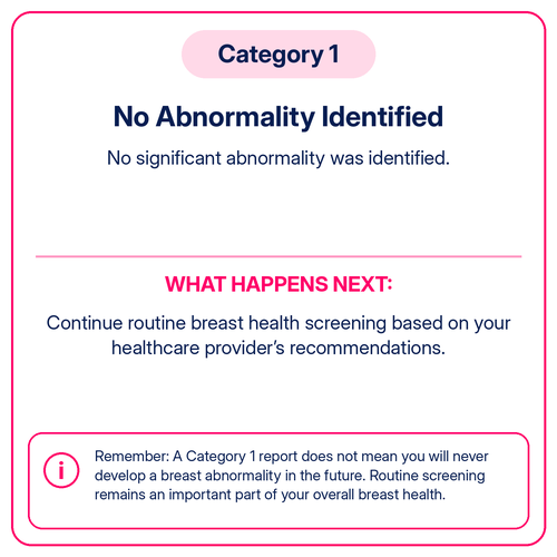
Your report category helps communicate what was seen during your screening and whether additional evaluation may be recommended.

Category 1
Category 2
Category 3
Category 4

How Findings Are Mapped and Measured

If your screening identifies an area that needs a closer look, your report may include details about its location and size. These measurements help your healthcare provider understand exactly where the finding was observed.

At HerScan, sonographers document findings using a clock-face system, the distance from the nipple, and the size of the area identified. This information is reviewed by a board-certified radiologist to help determine whether additional evaluation is recommended.
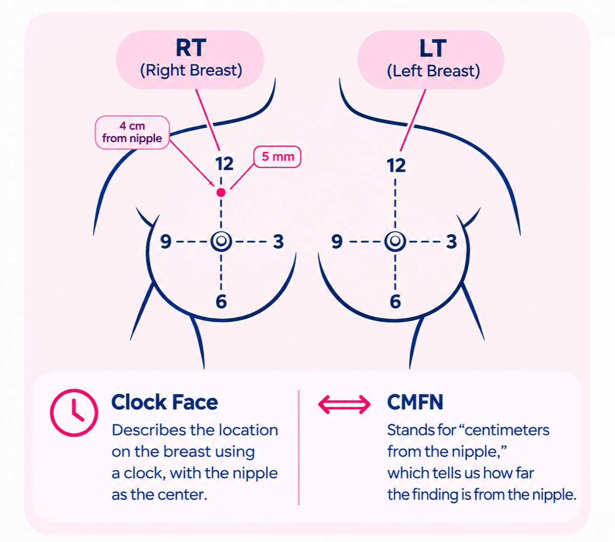
How findings are mapped and measured

Why Might Additional Breast Imaging Be Recommended?

  • Clarify a finding: A diagnostic exam can take a more focused look at a particular area.

  • Compare imaging methods: Mammography, ultrasound, and MRI show breast tissue differently.

  • Evaluate a specific area: Diagnostic imaging can focus specifically on the area identified during screening.

  • Monitor a finding: Some findings may be followed over time to evaluate whether they change.

  • Determine whether even further testing is needed: Depending on the results of diagnostic imaging, additional testing, such as a biopsy, may be recommended.

breast cancer ultrasound image

For example, on a mammogram, dense breast tissue can make some findings harder to see.

A Note About HerScan Categories and BI-RADS:

HerScan does not use BI-RADS risk assessment to assign an individual's probability of breast cancer. Your HerScan screening category communicates the findings and recommended next steps from your screening. It should not be interpreted as a diagnosis or as a specific percentage likelihood that cancer is present.

Questions About Your Results?

Contact Us →

Frequently Asked Questions

Please get in touch with us for all other inquiries: herscanresults@herscan.com.

What do the colors mean on the breast ultrasound when I’m getting screened?

Color Doppler is used to evaluate blood flow. The colors generally show whether blood is moving toward or away from the ultrasound probe. A common convention is BART: Blue Away, Red Toward. The colors themselves do not indicate whether something is cancerous or benign.

Yes. An abnormal screening result does not automatically mean breast cancer. Many findings that need a closer look are ultimately found to be benign. Additional diagnostic imaging may be recommended to better evaluate the area.

HerScan does not use BI-RADS to provide an individual breast cancer risk assessment. HerScan screening reports use result categories to communicate what was identified during the screening and what follow-up may be recommended. 

Screening is designed to look for abnormalities. If something is identified that needs a closer look, diagnostic imaging may be recommended to focus on that specific area, gather additional information, and help determine whether any further testing is needed.

No. A HerScan Category 4 result means an abnormality was identified that requires further diagnostic evaluation. It is not a breast cancer diagnosis. Your healthcare provider or breast imaging center can perform the additional evaluation needed to learn more about the finding.

Questions About Your Results?

Contact Us →