breast cancer screening
Dense Breast Tissue Ultrasound
Did you know that women with dense breasts are more likely to develop breast cancer than women with low density? (source)
Nearly half of women have dense breast tissue, which can make it harder to see certain findings on a mammogram. That’s why many women benefit from supplemental screening to get a more complete picture.
Breast ultrasound is a comfortable, compression-free and radiation-free way to screen breast tissue and the underarm (axilla) area for abnormalities.

Screens breast tissue & underarm region
A complete screening ultrasound surveys both breasts and the axilla to look for abnormalities, often before you can feel anything.
Helpful for dense breast tissue
Because density can mask cancers on mammography, ultrasound can add another way to evaluate tissue that may be harder to interpret on other modalities.
Compression free and generally comfortable
During your appointment, a probe glides over both breasts and the axilla area, without compression or discomfort.
Proactive screening if you have a family history
The goal of screenings is to detect breast cancer early, when it is most treatable.
High accuracy for implant surveillance
In studies, ultrasound showed ~90.9% sensitivity and 100% specificity for silicone implant rupture detection
No referral or doctor's order needed
Eliminating barriers to breast screenings is our mission. No prescription or referral from a doctor is required.
-1.png)
Dense Breast Screening Using Ultrasound Technology
Breast ultrasound uses sound waves (not radiation) to create real-time images of breast tissue and the underarm (axilla) region. It can help identify findings such as solid masses or fluid-filled cysts, and it’s commonly used both as supplemental screening and as follow-up imaging when something needs a closer look.
Who is Breast Ultrasound Screening For?
Have dense breasts and want supplemental screening
Are higher-than-average risk and want additional screening
Want to be proactive between routine screenings. Many breast changes can develop quietly, and screening is about catching concerns early
Want a radiation-free, compression-free screening experience that evaluates both breast tissue and the axilla region
Breast Cancer Rates Are Rising in Younger Women
Breast cancer still occurs most often at older ages—but multiple surveillance sources show increasing incidence in younger women
Our mission is to make breast ultrasound screening accessible to women everywhere, without insurance barriers or age restrictions
NIH analyses of U.S. cancer trends also show breast cancer is among the cancers with rising incidence in people under 50
When breast cancer is found earlier, treatment options are often less extensive and outcomes are better
Levels of breast density are categorized by using the American College of Radiology's Breast Imaging Reporting and Data System. This is also known as BI-RADS. These levels are scored from A to D and describe the level of density:
- Category A: The breast is made up of almost all fatty tissue (about 10% of women).
- Category B - There are scattered areas of dense glandular and fibrous tissue (about 40% of women).
- Category C - More of the breast is composed of dense glandular and fibrous tissue, also described as heterogeneously dense (about 40% of women).
- Category D - The breast is extremely dense, making it much more difficult to see masses on a mammogram (about 10% of women).
Generally speaking, if you receive a mammogram report
says your breasts are heterogeneously or extremely dense,
then you are considered to have dense breast tissue.
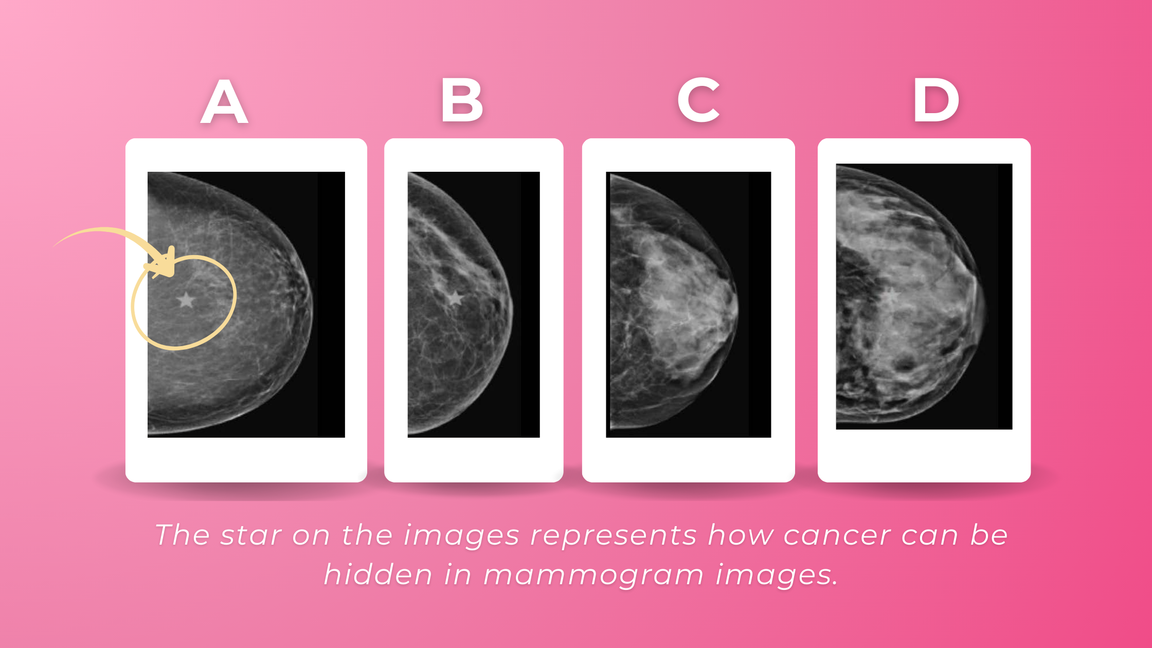
On a mammogram, fatty breast tissue appears dark, making tumors (white areas) easier to see. In dense breasts, both normal tissue and tumors look white, which can hide cancers and make them harder to detect.